Table of Contents
Calyx Of The Kidney
Calyx Of The Kidney – The outer whorl of the flower is known as the sepal. Sepals are the functional units of sepals, which means that sepals are a collection of sepals. The sepals are usually green in colour and protect the internal structures of the flower from breakage, mechanical damage and desiccation. It is compatible with other internal systems of the flowers, such as the corolla, the gynoecium and the androecium. The corolla is the set of petals, the androecium is the male reproductive flower, and the gynoecium is the female reproductive flower.
The cup is located below the corolla. In some plants, the calyx and corolla are indistinguishable and are called the perianth. Once the flower blooms, the calyx continues to support the development of the fruit. Occasionally an additional whorl is found outside the calyx consisting of a whorl of bracts arising from the union of the sepal appendages.
Calyx Of The Kidney – Forms of Calyx
Polysepal: When the calyx consists of the calyx devoid of each other, it is known as polysepalous. For example, Rose, Cassia
Gamosépalo: when the sepals fuses with the calyx, it is called a gamosepalous. For example, Datura
Expiration: When a flower withers or falls, it is called expiration. For example, poppy
Polaroid: Under petaloid conditions, the sepals of the flowers are colourful. For example, delphinium
Persistence: In this way, the sepals do not wither and persist even during fruiting. For example, the eggplant
Calyx Of The Kidney – Major Calyx
The Major calyx surrounds the top of the Malpighian pyramids. Urine in the kidney passes through the papilla at the apex into the calyx and then into the calyx before passing through the renal pelvis into the ureter.
Smooth muscle peristalsis arising in velocity cells in the cups’ walls pushes urine through the pelvis and ureters into the bladder.
Renal Calyx (Greater Calyx)
A cup not completely darkened on a urogram (phantom cup) is often a precursor of serious underlying kidney disease. Causes of the phantom cup include tuberculosis, tumours, stones, ischemia, trauma, and congenital anomalies. The pathologic basis of the radiographic findings in each of these entities is described, and a comprehensive approach to diagnosis is outlined.
Functions:
- It ensures the flower’s life as protection, photosynthesis and defence against insects.
- The main function of the sepals is to protect the flowering plant during the newborn stage.
- When mature, they support the flower.
Structure Of the Human Kidney
Kidney
The kidney, in vertebrates and some invertebrates, is an organ that maintains water balance and expels metabolic waste. The primitive and embryonic kidney comprises two series of specialized tubes that open into two collecting ducts, the Wolffian ducts (see Wolffian vent). The most advanced kidney (metanephros) of adult reptiles, birds, and mammals is a compact organ associated with functional units called nephrons that filters primary urine from the blood, reabsorbs water and nutrients, excretes waste products, and produces final urine. , which is excreted.
The kidneys of reptiles and birds consist of many small lobes, which in birds fuses into three or more lobes. The collecting ducts empty in each lobe into a separate branch of the ureter. Thus, reptiles have relatively few nephrons (3,000 to 30,000 in lizards), whereas birds have large numbers (about 200,000 in birds, twice as many as mammals of similar size).
Description
Mammalian kidneys have a somewhat granular outer section (the cortex), which contains the glomeruli and convoluted tubules, and a smooth, rather rough inner section (the medulla), which includes the loops of Henle and collecting ducts. And also, as the ureter enters the kidney, it expands into the lumen, the renal pelvis. Urine passes into this container from the collection tubes—numerous nephrons (20,000 in a rat).
Human kidneys are about 10 centimetres long and are located below the diaphragm and behind the peritoneum.
Therefore, each kidney contains 1,000,000 to 1,250,000 nephrons that filter the five litres of water from the blood every 45 minutes, equivalent to 160 litres per day. Of this, only 1 1/2 litres come out; The nephron absorbs the rest.
Damaged kidneys secrete an enzyme called renin, which stimulates blood vessels to constrict. When the damage is initially due to high blood pressure, the increased pressure in the narrowed vessels leads to further kidney damage.
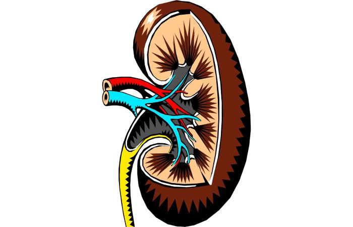
Calyx Of The Kidney – Internal Structure
- Viewed from the inside, the kidney contains an outer layer of the cortex that surrounds the inner medulla.
- The pulp consists of a series of medullary pyramids named for their triangular shape. They scratch because they contain microscopic coiled tubes called nephrons, the kidney’s functional unit.
- Therefore, the nephrons produce urine and empty it into small collecting ducts within the medullary pyramids. The collecting ducts fuse at the base of the pyramids to form the renal papilla.
- From the papilla, urine flows into cup-shaped structures called the major and minor calyces. From
- the calyces, urine flows into the larger open space of the renal pelvis, acting like a funnel that drains urine from the kidney into the ureter.
Calyx Of The Kidney – Blood Flow
- Blood flows to the kidneys through the right and left renal arteries. In each kidney, these branches develop into smaller arterioles.
- Blood is under very high pressure and flows through the arteries to a small knot of vessels called the glomerulus. These are found in the nephrons.
- And also, blood pressure from the glomerulus decreases, and blood flows into the arterioles surrounding the nephrons. These, in turn, connect to a series of small veins, and these vessels unite and eventually form the renal vein.
- Thus, about a quarter of the total cardiac output (or total blood flow) circulates in the kidneys, equating to just over a litre of blood per minute.
Why Are Kidney Stones Form?
Kidney stones form for many reasons. For example, you won’t have enough urine to dilute the chemicals if you don’t drink enough water. The substances can then form crystals that can turn into stones. Here are some of the causes of kidney stones:
- Loss of fluids (dehydration). This can cause urine to become concentrated, resulting in stone formation.
- Certain foods. Some foods contain large amounts of chemicals that sometimes crystallize into stones. Eating foods that have a lot of meat or salt can make kidney stones worse.
- Kidney infections. These infections promote stones by slowing the flow of urine or altering the urine’s acid balance.
- Family history: If family members have had kidney stones, you are more likely to have them too.
- Deficiency of certain substances in the urine. Certain materials can protect you from the stone formation. The stone formation can increase if you don’t have enough in your urine.
Conclusion
In this article, we get fettle explains Calyx Of The Kidney. We discuss the Forms of Calyx, the Structure of the kidney, and more. Thus, the above details are just for informational purpose.
Helpful Resources:
High-Fiber Rich Foods You Should Be Eating